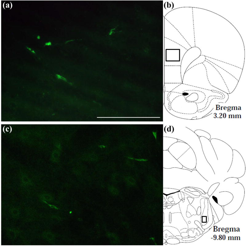
Fig. 6.
Localization of a green-fluorescent tagged OxA peptide in the brain after intranasal administration. (a) Typical distribution of the fluorescent-tagged OxA peptide (50 µl, 500 µM) in the PrLC after intranasal administration. Green fluorescence indicates the appearance of the labeled peptide in the PrLC. (b) Schematic indicating the approximate location (black-outlined square) within the PrLC where fluorescence photomicrographs were obtained. (c) Typical distribution of the fluorescent-tagged OxA peptide in the SpTrN. (d) Schematic indicating the approximate location (black-outlined square) within the SpTrN where fluorescence photomicrographs were obtained. Abbreviations: OxA, orexin-A; PrLC, prelimbic prefrontal cortex; SpTrN, spinal trigeminal nucleus. Scale bar approximately 100 µm.

