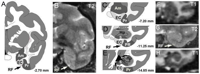
Figure 2. Location of entorhinal cortex (EC) in rhesus monkey with corresponding MR images.
Schematic diagrams (adopted from Paxinos et al., 1989) of entorhinal cortex across four coronal planes from −2.7 to −14.85 mm relative to bregma (A, C–E). The entorhinal cortex is located on the ventral and medial surface of the temporal lobe, with local landmarks that include the rhinal fissure (RF), perirhinal cortex (PR), amygdala (Am), subicular area (S), and hippocampus (Hp). T1 and T2 MRI scans (B, F–H) with the visible landmark of the rhinal fissure (RF) indicated on the T2 image (arrows in A,B,D,G).

