
Figure 3. Colocalization of NO and SNO-NP fluorescence in lymph nodes (LN) draining site of injection demonstrates nanoparticle-mediated lymphatic transport facilitates NO delivery to LN.
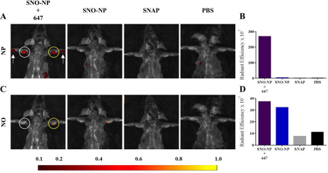
Representative IVIS images (A, C) and quantification (B, D) of AlexaFluor647 (A–B) and NO probe DAF-FM DA (C–D) fluorescence in draining LN associated with SNO-NP, but not SNAP administration in the forelimb skin. White arrow, intradermal site of treatment group injection; white circle, LN draining the site of intradermal injection; yellow circle, DAF-FM DA injected LN.