Figure 7.
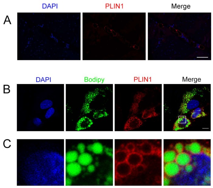
PLIN1 localized in porcine longissimus dorsi muscle and adipocytes. (A) Immunohistochemistry of PLIN1 in porcine longissimus dorsi muscle. PLIN1 was stained with antibody PLIN1 (red). Nuclei were stained with 4′,6-diamidino-2-phenylindole (DAPI, blue). Scale bars: 100 μm; (B,C) PLIN1 immunofluorescence at the surface of lipid droplets (LDs) in porcine adipocytes. PLIN1 was stained with antibody PLIN1 (red). Lipid droplets were stained with Bodipy (green). Nuclei were stained with DAPI (blue). Scale bars: 10 μm.

