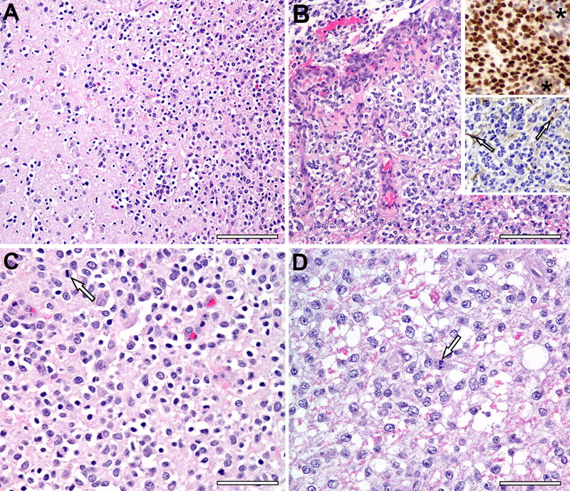
Figure 1. Histology of Canine Glioma Samples.
A) Grade III oligodendroglioma (upper right) invades adjacent cerebral parenchyma. Scale bar, 100 µm. (B) Microvascular proliferation is prominent around a necrotic focus (top) in Grade III oligodendroglioma. Scale bar, 100 µm. Upper inset depicts Olig2 immunohistochemistry with nuclear labeling in neoplastic cells but no Olig2 expression by cells of the microvascular proliferation (asterisks). Lower inset depicts processes of a few GFAP–positive astrocytes (arrows), but the neoplastic cells are negative. (C) Most neoplastic cells in this Grade III oligodendroglioma have a hyperchromatic nucleus with occasional mitotic figures (arrow) and a clear perinuclear ‘halo’, an artifact of delayed fixation. Scale bar, 50 µm. (D) Neoplastic cells in this Grade III oligodendroglioma have a larger hypochromatic nucleus with occasional mitotic figures (arrow) and pale eosinophilic cytoplasm with indistinct cell borders. Scale bar, 50 µm. (n = 10)

