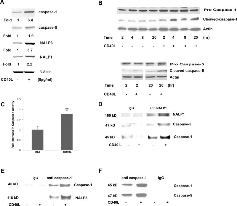
Fig 3.
CD40L-induced inflammasome activation in hRPE cells. mRNA levels of inflammasome components caspase-1, caspase-5, NALP1 and NALP3 were determined by RT-PCR in hRPE cells treated with or without CD40L for 6 hr (A). The fold changes were calculated by normalization against β actin and comparison with untreated control mRNA levels. Caspase-1 and -5 protein cleavage in hRPE cells was analyzed by Western blot with or without CD40L for 2, 4, 8, or 20 hr (B). An antibody against actin was used in each Western blot analysis as a loading control. Untreated (Ctrl) and CD40L treated hRPE cell lysate were examined for caspase-1 activation by a caspase-1 assay kit (C). Co-IP pulldown of anti-NALP1 (D) or anti-caspase-1 (E, F) followed by Western blotting using antibodies against NALP3, caspase-1, NALP1 or caspase-5. Anti-IgG antibody was used as a control for immunoprecipitation. All concentrations of CD40L in these experiments were 5µg/ml. **P<0.01 as compared with untreated control (C).

