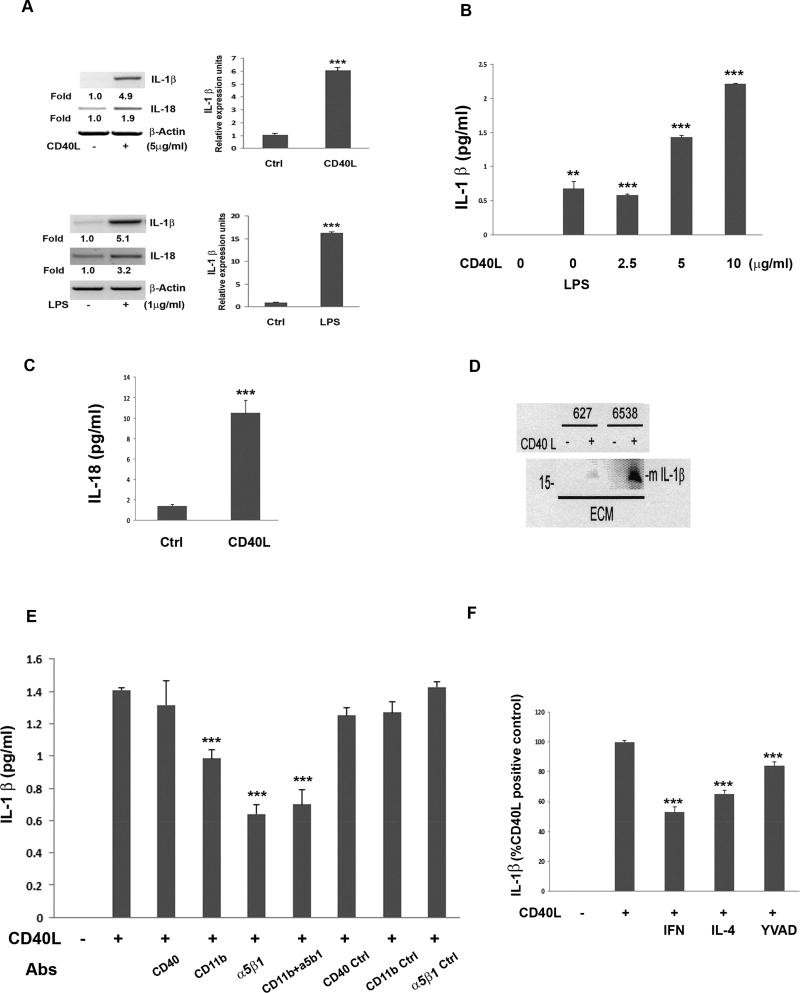
Fig 4.
Detection of CD40L-induced cleaved, mature IL-1β from hRPE cells through CD40L receptors CD11b and α5β1. mRNA levels measured by RT-PCR and qPCR of IL-1β and IL-18 with or without CD40L (5 ug/ml) or LPS (1 µg/ml) for 6 hr (A). IL-1β secretion from hRPE cells cultured with CD40L (0, 2.5, 5, 10 µg/ml) or LPS (1 µg/ml) for 24 hr (B). CD40L-induced hRPE secretion of IL-18 (C). hRPE cells cultured from 2 donors (627 and 6538) were left unstimulated or stimulated with CD40L followed by Western blot analysis for cleaved IL-1β in the extracellular media (ECM)(D). IL-1β ELISA of conditioned media from hRPE cells cultured with or without CD40L for 24 hr in the presence or absence of antibodies (Abs) against CD40 (5 µg/ml), CD11b (20 µg/ml) or α5β1 (10 µl/m), as well as the corresponding isotype control (E). hRPE cells were cultured with or without CD40L in the presence or absence of IFN-γ (IFN, 500 U/ml), IL-4 (100 ng/ml), or Z-YVAD-FMK (YVAD, 2µM) for 24 hr (F). The concentrations of CD40L were 5 µg/ml (C–F). *p<0.05; **p<0.01; ***p<0.001, as compared with untreated control (A–C) or CD40L-stimulated control (E, F). Ctrl, Control; LPS, lipopolysaccharide; Abs, antibodies.

