
Figure 1.
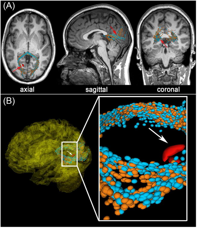
Representative example of DTI streamlines passing through the vicinity of a ∼4 mm3 CMB (red) in an old adult victim of mTBI. Arrows indicate a CMB in the left hemisphere, close to a streamline bundle belonging to the splenium of the corpus callosum. (A) Standard views (coronal, sagittal, axial) of T1-weighted MRI are shown in addition to DTI glyphs associated with perilesional WM streamline bundles imaged acutely (orange) and approximately six months after injury (light blue). The splenium is notably asymmetric at both time points, with the asymmetry being most pronounced close to the CMB (inset). (B) Splenial streamlines ipsilateral to the CMB diverge briefly in its vicinity, and this is not found to occur contralateral to the CMB (inset). This asymmetry is also found at the time of the chronic scan.