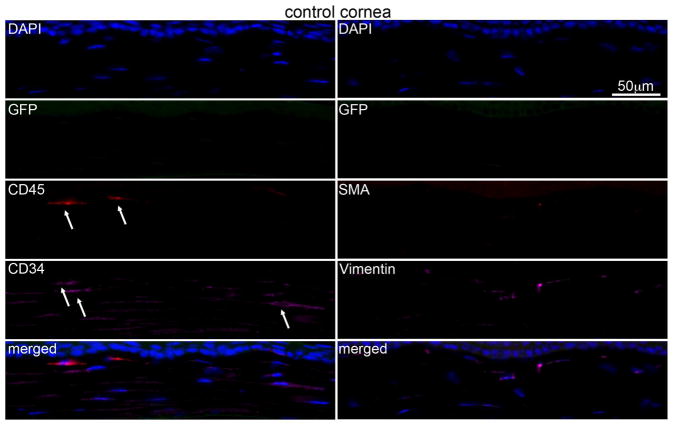
Fig. 2.
Multiplex immunohistochemistry (IHC) in the chimeric unwounded control mouse corneas. No visible bone marrow derived GFP or αSMA positive cells were detected in any unwounded control corneas. Few CD45+ (arrows) and/or CD34+ (arrows) cells were present in control corneas and these cells were GFP-negative. Blue is DAPI staining of cell nuclei and the merged panel is an overlay of all the images in that column. Negative control IHC was performed for all antigens without primary antibody and no staining was detected (see supplementary Fig. S1). 400x mag.

