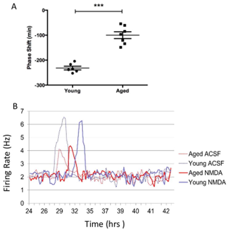
Figure 4.

Cellular firing rhythms in SCN brain slices from young and aged mice. (A) Electrophysiological recordings from isolated aged SCN brain slices displayed attenuated shifts in the peak firing rhythms following exposure to NMDA in comparison with preparations from younger animals (p<0.001, n= 6 young mice; n= 7 aged mice). (B) There was also an observed decrease in the peak amplitude of firing in the aged samples (n=7; 4.07 ± 0.21 Hz (mean ± SEM)) relative to recordings taken from young mice (n=6; 6.02 ± 0.15 Hz, p <0.01). Here we show the frequency of SCN cells firing rates represented by a 1-h running mean with a 15-min lag over time for four individual slices recorded from young and aged mice.
