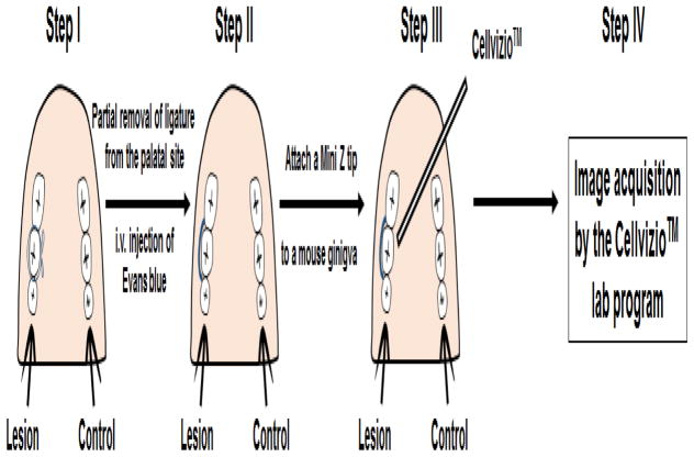
Figure 2.
Schematic illustration of developed intravital endoscopic imaging of experimental periodontal lesions in a mouse model of periodontitis using the Cellvizio™ device. Step I: Induce experimental periodontitis by placing a ligature at the second maxillary molar; Step II: Partially remove ligature at the palatal site and inject Evans blue solution i.v.; Step III: Attach a mini Z tip directly to the gingival tissue and image; Step IV: Collect data.

