
Figure 5. The TRIF-dependent effector cytokines IFN-β and IL-1β augment the Tfh and antibody responses to the killed vaccine.
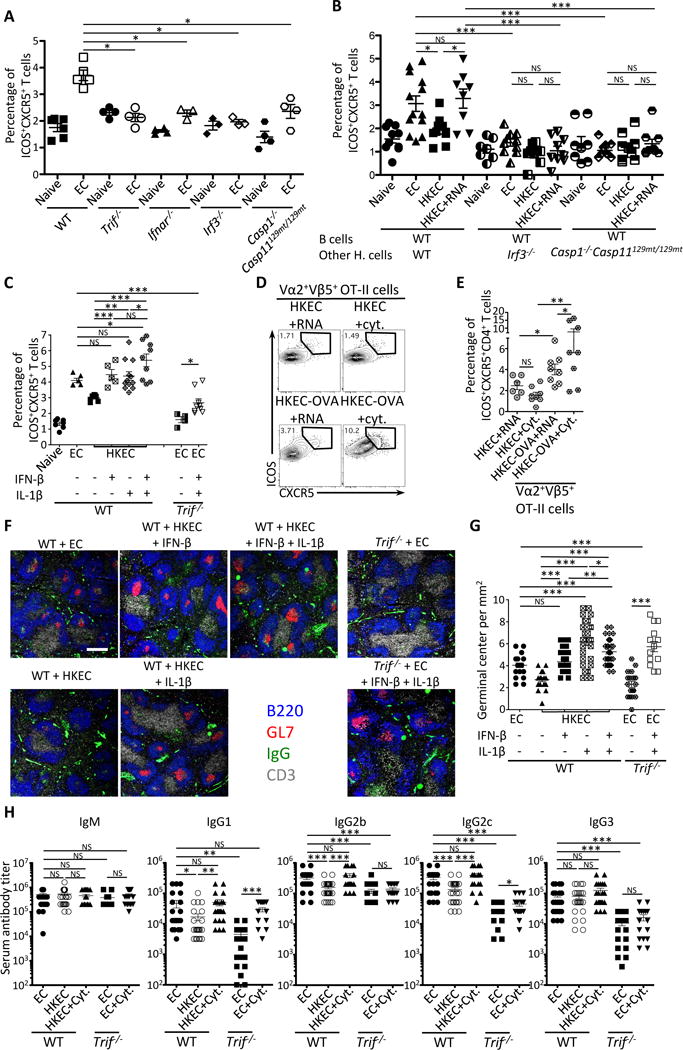
(A) Percentages of CXCR5+ICOS+CD4+ T cells in naïve and live EC vaccinated mice indicated on the X-axis.
(B) Percentages of CXCR5+ICOS+CD4+ T cells in indicated naïve or vaccinated chimeric mice generated as in Figure 4A, except here lethally irradiated mice received 20% B cell-sufficient WT BM and 80% B cell-deficient μMT BM on either WT, Irf3−/− or Casp1−/−Casp129mt/129mt backgrounds.
(C) Percentages of ICOS+CXCR5+CD4+ T cells in WT or Trif−/− mice that had received indicated vaccines.
(D, E) Flow cytometry dot plots (D) and percentages (E) of CXCR5+ICOS+ cells within gated Vα2+Vβ5+ transgenic OT-II TCR expressing CD4+ T cells adoptively transferred into mice 48 hours before receiving indicated vaccines.
(F, G) Immunofluorescence micrographs at 4X magnification on spleen sections from indicated mice stained for B220, GL-7, IgG and CD3. Scale bar = 300μm (F), and quantification of GC per mm2 (G) where each symbol represents an individual field of view.
(H) Day 25 serum titers of class-specific anti-EC antibodies.
Tfh and GC responses were measured in spleens on days 5 and 7, respectively, after vaccination of indicated mice with 5×10⁷ live EC, HKEC or HKEC+RNA(30μg). 50 U IFN-β and/or 1μg IL-1β were injected intravenously 20 hours after vaccination.
NS, not significant (P > 0.05); *, P<0.05; **, P≤0.01 and ***, P≤0.001 (two-tailed unpaired t test). Data are mean±s.e.m. Numbers adjacent to outlined areas indicate percent of cells in gates. Mouse numbers are in (A) WT(naive, n=6 ;+EC, n=5), Trif−/−(naive, n=4 ;+EC, n=4), Ifnar−/−(naive, n=4 ;+EC, n=3), Irf3−/−(naive, n=3 ;+EC, n=3) Casp1−/−Casp11129mt/129mt(naive, n=4 ;+EC, n=4); (B) WT(WT B cells)(naive, n=10 ;+EC, n=13; +HKEC, n=10; +HKEC+RNA, n=8), Irf3−/−(WT B cells)(naive, n=7 ;+EC, n=10; +HKEC, n=9; +HKEC+RNA, n=9), Casp1−/−Casp11129mt/129mt(WT B cells)(naive, n=8 ;+EC, n=10; +HKEC, n=9; +HKEC+RNA, n=9); (C) WT(naive, n=7 ; +EC, n=6; +HKEC, n=7; +HKEC+IFN-β, n=5; +HKEC+IL-1β, n=12; +HKEC+IFN-β+IL-1β, n=9) and Trif−/−(+EC, n=4; +EC+IFN-β+IL-1β, n=9); (E) WT+OTII T cells(+HKEC+RNA, n=6; +HKEC+IFN-β+IL-1β, n=8+HKEC+RNA, n=8; +HKEC+IFN-β+IL-1β, n=8); (G) WT(+EC, n=5; +HKEC, n=4; +HKEC+IFN-β, n=6; +HKEC+IL-1β, n=6; +HKEC+IFN-β+IL-1β, n=6) and Trif−/− (+EC, n=3; +EC+IFN-β+IL-1β, n=4); (H) WT(+EC, n=25; +HKEC, n=28; +HKEC+IFN-β+IL-1β, n=23) and Trif (+EC, n=22; +HKEC, n=15; +HKEC+IFN-β+IL-1β, n=18).
See also Figure S5.