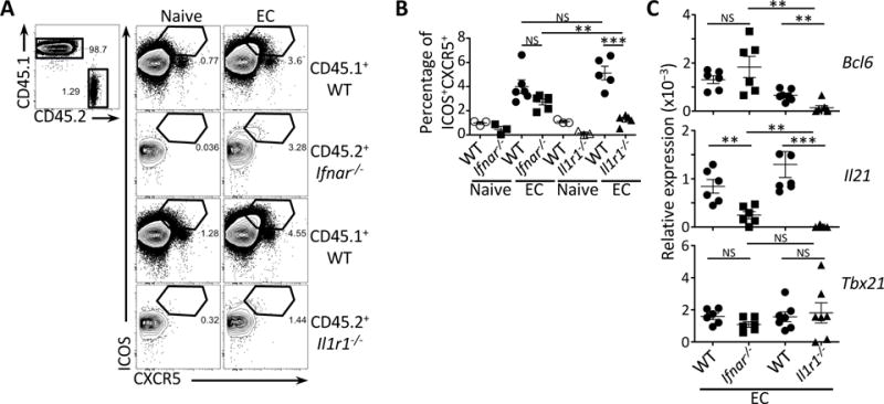
Figure 6. T cell intrinsic role of TRIF-dependent cytokines in Tfh cell differentiation.

(A) Flow cytometry dot plots for ICOS and CXCR5 on CD45.1+ or CD45.2+CD4+ T cells that were either Ifnar−/− or Il1r1−/−.
(B) Percentages of ICOS+CXCR5+CD4+ T cells within the CD45.1+ or CD45.2+ populations from (A).
(C) Quantitative RT-PCR analysis for Bcl6, Il21 and Tbx21 transcripts in sorted CD44+CD4+ T cells either CD45.1+ (WT) or CD45.2+ (Ifnar−/− or Il1r1−/−). Data represent expression relative to β-actin.
(A, B, C) Data on pre-gated CD4+ T cells showing endogenous CD45.1+ cells or adoptively transferred CD45.2+ (Ifnar−/− or Il1r1−/−) CD4+ T cells isolated from spleens 5 days after injection of 5×10⁷ live EC.
NS, not significant (P > 0.05); *, P<0.05; **, P≤0.01 and ***, P≤0.001 (two-tailed unpaired t test). Data are mean±s.e.m. Numbers adjacent to outlined areas indicate percent of cells in gates. Mouse numbers are in (B) WT+ Ifnar−/− T cells (naive, n= 3; +EC, n=6) and WT+ Il1r1−/− T cells (naive, n= 3; +EC, n=5); (C) WT+ Ifnar−/− T cells (+EC, n=6) and WT+ Il1r1−/− T cells (+EC, n=7).
See also Figure S6.
