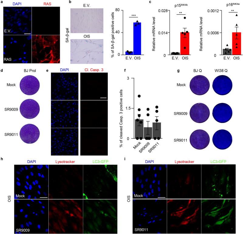
Extended Data Figure 9. REV-ERBs agonists do not affect viability of normal proliferating and quiescent cells OIS.
a, Immunofluorescence assay for RAS confirms RAS overexpression in OIS cells. b, SA-β-Gal assay shows induction of senescence (n=3 biological independent samples Student's t-test one-tailed ****P<0.0001). c, Induction of cell cycle inhibitors p15INK4b, p16INK4a is assayed by qRT-PCR; n=5 independent biological samples; Mann–Whitney test one-tailed p15INK4b **P =0.004, p16INK4a **P=0.0079. d–e, REV-ERBs agonists’ do not induce apoptosis in proliferating and quiescent normal diploid fibroblasts BJ (d–g), as shown by proliferation assay (d, 7 days, 20µM) and immunofluorescence for cleaved Caspase 3 (e–f, 7 days 20µM, one-way ANOVA; n=biological independent samples, n=7 (mock) n=5 (SR9009, SR9011) P=ns. Cell viability is also not affected in an additional normal diploid cell line WI38 (g, 10 days 20µM). h–I, SR9009 and SR9011 inhibit autophagy in OIS cells as shown by massive accumulation of lysosomes (lysotracker) and absence of LC3 puncta (3 days 20µM). All scale bars 50 µm. Data in a–i are representative of three independent experiments with similar results unless otherwise specified. All the data are plotted as mean ± s.e.m.

