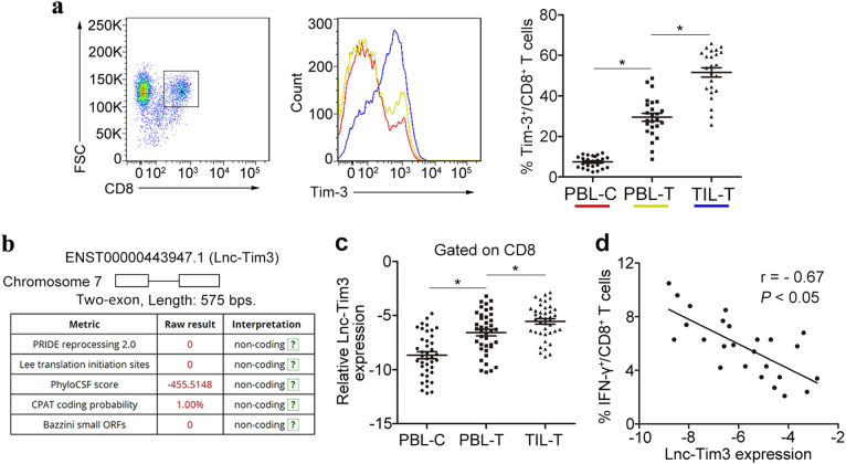
Fig. 1. Upregulated Lnc-Tim3 correlates with the exhaustion of CD8 T lymphocytes.
a The percentage of Tim-3+ CD8 T cells in the infiltrated CD8 T cells was determined by flow cytometry. The percentages of Tim-3+ CD8 T cells in the tumor-infiltrating CD8 T cells (TIL-T) and peripheral blood CD8 T cells of HCC patients (PBL-T) and healthy controls (PBL-C) was analyzed. n = 25 for each group. b The protein-coding potential and the ORF size (a two-exon gene) of long non-coding RNA Lnc-Tim3 (ENST00000443947.1) were analyzed by databases. c Relatively increased levels of Lnc-Tim3 were confirmed in tumor-infiltrating CD8 T cells (TIL-T) by comparing them with those in the peripheral blood CD8 T cells of HCC patients (PBL-T) and healthy controls (PBL-C). n = 40 for each group. d The Pearson’s correlation of Lnc-Tim3 expression and the percentage of IFN-γ+ CD8 T cells in the tumor-infiltrating CD8 T cells was analyzed. (n = 25). Data are presented as means ± S.E.M. (*P < 0.05)

