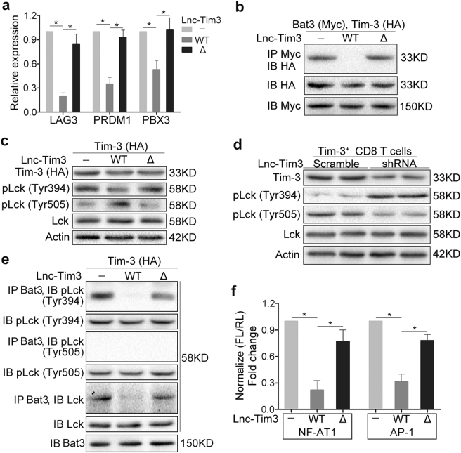
Fig. 4. Upregulated Lnc-Tim3 in CD8 T cells induces an exhausted-like phenotype via suppressing Tim-3-Bat3 signaling.
a CD8 T cells isolated from peripheral blood of healthy human were transfected with indicated lentiviral particles (wild type Tim-3 and Lnc-Tim3 wild type or Δ226-277). The mRNA levels of LAG3, PRDM1, and PBX3 were assessed by real-time PCR after cells treated with anti-TCR/anti-CD28 beads and PMA-ionomycin. (n = 10). b A western blot of Bat3-Myc, Tim-3–HA, and Lnc-Tim3 (wild type or Δ226-277) overexpressed in HEK 293 T cells and coimmunoprecipitated to confirm the specificity of the Bat3–Tim-3 interaction in mammalian cells. IP, immunoprecipitation; IB, immunoblot. Representative of three experiments. c A western blot of pLck (Tyr394 and Tyr505) expression from Jurkat T cells that were transfected with indicated lentiviral particles (Tim-3–HA, Lnc-Tim3 wild type or Δ226-277) and anti-TCR/anti-CD28 beads. Representative of three experiments. (d) Lnc-Tim3highTim-3+CD8 T cells isolated from tumor-infiltrating lymphocytes (TILs) of HCC patients with Lnc-Tim3 high expression were transfected with indicated lentiviral particles (Lnc-Tim3 scramble or shRNA) and treated with anti-TCR/anti-CD28 beads. The expression of pLck (Tyr394 and Tyr505) was assessed by western blot. Representative of three experiments. e Jurkat T cells were transfected with indicated lentiviral particles (Tim-3–HA, Lnc-Tim3 wild type or Δ226-277) and treated with anti-TCR/anti-CD28 beads and PMA-ionomycin, followed by coimmunoprecipitated to confirm the specificity of the Bat3-pLck (Tyr394 and Tyr505) interaction in cells. Representative of three experiments. f Jurkat T cells were transfected with indicated lentiviral particles (Tim-3–HA, Lnc-Tim3 wild type or Δ226-277). Then, Jurkat T cells were also transfected with NFAT1 or AP-1 luciferase reporter. NFAT1 or AP-1 luciferase activity was determined after a 6-h stimulation (anti-TCR/anti-CD28 beads and PMA-ionomycin). Data are presented as means ± S.E.M. (*P < 0.05)

