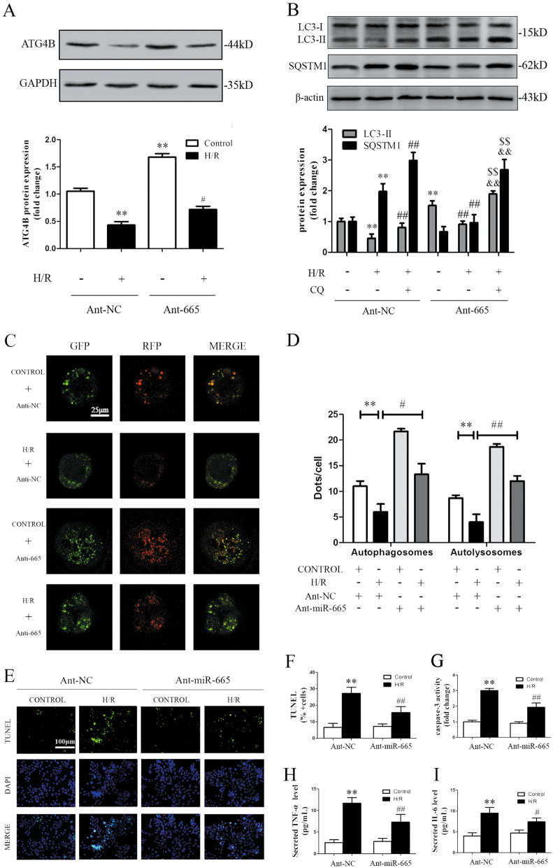
Fig. 6. Inhibition of miR-665 stimulates autophagic flux and mitigates cell apoptosis and inflammation in Caco-2 cells treated with H/R.
a Ant-665 stimulated ATG4B expression in response to H/R. b Representative immunoblots of endogenous LC3B-II and SQSTM1 in Caco-2 cells that were transfected with Ant-NC or Ant-665 and subjected to H/R (n = 3). c Representative images showing LC3 staining in different groups of Caco-2 cells that were infected with the mRFP-GFP-LC3 adenovirus for 24 h. d Quantitative analysis of autophagosome and autolysosome formation (n = 3). e TUNEL and DAPI staining. f The percentage of apoptotic cells is represented as TUNEL-positive cells/DAPI (n = 6). g Caspase-3 activity (n = 6). h, i Secreted TNF-α and IL-6 in the cell culture supernatant were quantified by ELISA (n = 6). **P < 0.01 versus the control, #P < 0.05, ##P < 0.01 versus H/R, &&P < 0.01 versus H/R+CQ, $$P < 0.01 versus H/R + Ant-665

