
Figure 1.
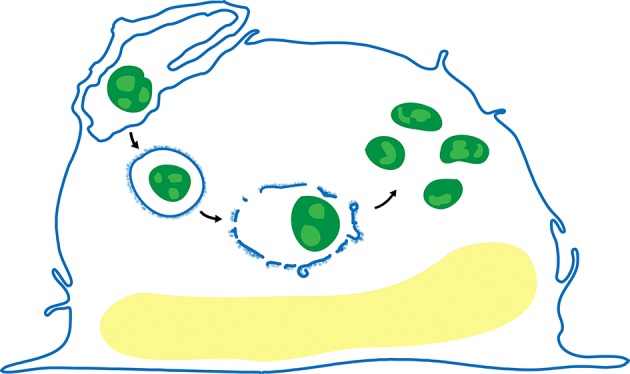
Life cycle of F. tularensis in human macrophages. After uptake by looping phagocytosis (upper left), the bacteria (green) reside in a membrane bound vacuole that often acquires a densely staining fibrillar coat (post first arrow), which subsequently forms blebs and vesicles (post second arrow), and disintegrates. The bacteria escape the phagosome and replicate freely in the cytosol (post third arrow).