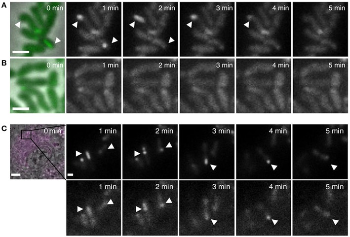
Figure 6.
(A,B) Live fluorescence microscopy shows dynamic formation and disassembly of IglA-sfGFP fluorescent structures in wild-type F. novicida (A) but not ΔpdpB F. novicida (B). Arrow heads indicate positions of fluorescent sheath assembly, contraction, and disassembly. Reproduced with permission from Brodmann et al. (2017). (C) Time-lapse images of unprimed wild-type BMDMs infected with F. novicida expressing IglA-sfGFP ClpB-mCherry2 for 1 h. First image shows merged phase contrast, GFP and mCherry channels with a 30 × 30 μm field of view. Scale bar, 5 μm. Close ups show GFP channel (upper panels) and mCherry channel (lower panels). Close ups show 5 × 5 μm fields of view. Scale bar, 1 μm. Arrowheads indicate positions of T6SS sheath assembly, contraction and location of sheath after contraction. Reproduced with permission from Brodmann et al. (2017).

