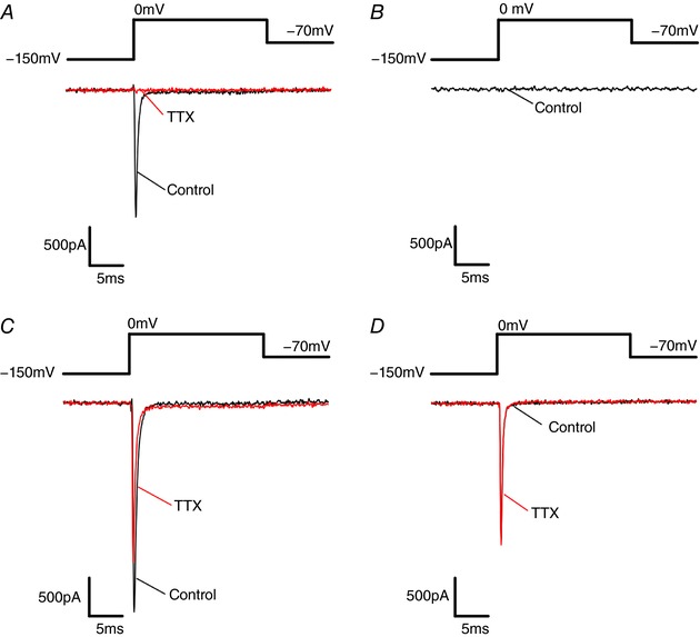
Figure 2. Endogenous NaV and mutant TTX‐resistant NaV1.7 currents in Ins1 and HEK cells.

A, endogenous NaV current from Ins1 cells in response to a step depolarization from −150 to 0 mV in the absence (black; control) and presence of TTX (red; TTX treated). B, no endogenous NaV currents were evoked in HEK cells by a step depolarization from −150 to 0 mV. C, NaV currents evoked in response to a step depolarization from −150 to 0 mV in Ins1 cells co‐transfected with mutant TTX‐resistant NaV1.7 and β1‐ and β2‐subunits in the absence (black; control) and presence of TTX (red; TTX treated). The reduction of the peak current induced by TTX reflects block of endogenous NaV current. D, same as in C but expressed in HEK cells. [Color figure can be viewed at http://wileyonlinelibrary.com]
