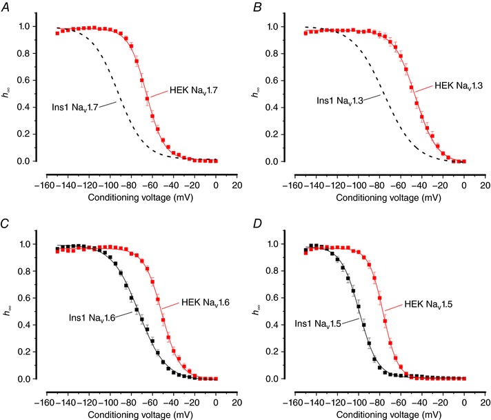
Figure 4. Voltage dependence of inactivation of NaV1.7, NaV1.3, NaV1.6 and NaV1.5 currents in Ins1 cells compared to HEK cells.

A, voltage dependence of NaV1.7 current inactivation (h ∞) when the α‐subunit is co‐expressed with β1‐ and β2‐subunits in Ins1 (same data as in Fig. 3 A: dashed curve) and HEK cells (red; n = 9). The curve represents a single Boltzmann fit to the data. B, as in A but for NaV1.3 (dashed curve same data as in Fig. 3 A; red, n = 7). C, as in A but for NaV1.6 (black, n = 35; red, n = 10). D, as in A but for NaV1.5 (black, n = 9; red, n = 10). See also Table 1. [Color figure can be viewed at http://wileyonlinelibrary.com]
