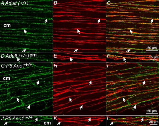
Figure 2. Ano1+ ICC‐IM are closely apposed to vAChT+ nerve fibres in the gastric fundus.

A–C, varicose VAChT+ nerve fibres (A, arrows, green) and Ano1+ ICC‐IM (B, arrows, red) within the circular layer (cm) of adult (+/+) fundus muscles. C, merged image showing the close apposition between vAChT+ nerve fibres and Ano1+ ICC‐IM (arrows). D–F, higher power images of the relationship between vAChT+ nerves and Ano1+ ICC‐IM. vAChT+ nerves were closely apposed to Ano1+ ICC‐IM for >250 μm. G–I, gastric fundus muscles of P5 Ano1+/+ animals also display a similar close morphological relationship between vAChT+ nerves and Ano1+ ICC‐IM as seen in adult animals. G, vAChT+ nerve fibres (arrows, green) and Ano1+ ICC‐IM (H, arrows, red) in the circular layer (cm). I, similar to adult tissues, vAChT+ nerves were closely apposed to Ano1+ ICC‐IM for >250 μm. J–L, at higher magnification, the close apposition between vAChT+ nerve fibres and Ano1+ ICC‐IM can be readily observed. Scale bars for each series of confocal reconstructions are shown in (C), (F), (I) and (L).
