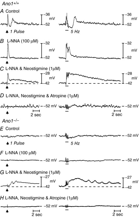
Figure 7. Nerve evoked post junctional responses in P5 Ano1+/+ and Ano1−/− mutant fundus muscles in response to EFS.

A, neural responses in Ano1+/+ fundus muscles under control conditions in response to EFS, 1 Hz (delivered at arrow, left) and 5 Hz (horizontal bar, right) respectively (n = 8). EFS evoked a frequency‐dependent biphasic motor response consisting of an initial fast transient EJP followed by a more sustained IJP that was often followed by a secondary depolarization in membrane potential before it returned to pre‐stimulus levels. B, in the presence of l‐NNA (100 μm), the EFS evoked IJP was abolished and the EJP increased in amplitude. C, in the continued presence of l‐NNA, neostigmine (1 μm) depolarized membrane potential, evoked an EJP and revealed a slower developing and more sustained depolarization in membrane potential (dashed lines) to EFS. D, atropine (1 μm) abolished both the initial and sustained depolarization at all EFS frequencies examined. E, in Ano1−/− mutants, EFS evoked little or no post‐junctional responses to EFS under control conditions at 1 Hz and a small hyperpolarization at 5 Hz (n = 15). F, l‐NNA (100 μm) produced little change to EFS at 1 pulse but attenuated the slight hyperpolarization in membrane potential at 5 Hz. G, in the continued presence of l‐NNA, neostigmine (1 μm) caused membrane depolarization and produced a slowly developing and sustained depolarization response to EFS, as in Ano1+/+ animals. H, atropine repolarized membrane potential and blocked the nerve evoked depolarization observed in the presence of neostigmine at all frequencies tested. Note also the difference in basal electrical activity between Ano1+/+ and Ano1−/− mutants.
