
Figure 1.
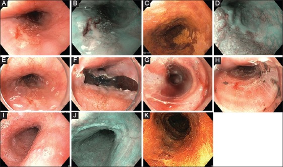
Endoscopic submucosal dissection of an early squamous cell carcinoma (T1am2). (A) High-definition white-light endoscopy visualization of a Paris 0-IIb lesion of the mid esophagus. (B) Narrow-band imaging and (C) Lugol coloration showing the limits of the lesion. (D) Narrow-band imaging with magnification showing the type V-2 intrapapillary capillary loops suggesting m2, resectable lesion. (E) Circular markings before endoscopic submucosal dissection. (F) Distal incision. (G) Submucosal dissection using the tunnel technique under the lesion. (H) Resection wound after en bloc endoscopic submucosal dissection. (I, J, K) Three-month follow-up endoscopy showing a clean esophageal, Lugol-negative scar, without evidence for recurrence or residual neoplasia