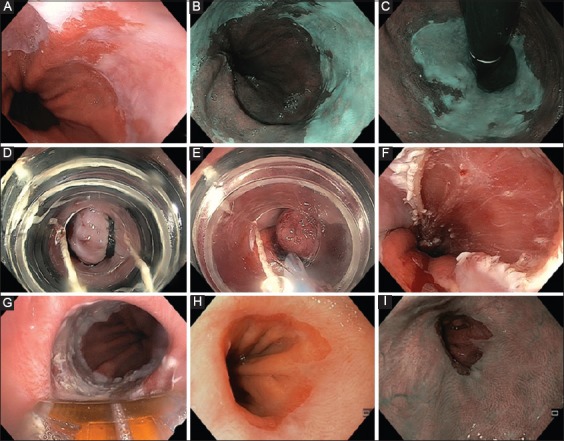
Figure 2.

Endoscopic treatment of early Barrett's neoplasia (T1am1). (A) High-definition white-light endoscopy showing a visible abnormality with nodularity and irregular nodularity and irregular pit pattern on a short Barrett's tongue. (B and C) Narrow-band imaging of the lesion in direct and retroflex view. (D) Band ligation of the lesion without submucosal lifting, before (E) placement of the snare below the band, and (F) resection wound after multiband mucosectomy. (G) Radiofrequency ablation using a focal probe to ablate residual Barrett's esophagus, 3 months after endoscopic mucosal resection. (H and I) Follow-up endoscopy 3 months later, showing a normal-appearing neo-Z line under white-light endoscopy (H) and narrow-band imaging (I)
