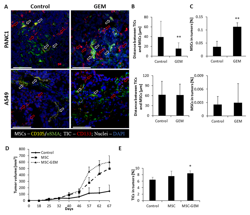
Figure 1. Chemotherapy induces the homing of MSCs to TICs in pancreatic tumors:
(A-C) Eight-to-ten week old SCID mice were subcutaneously implanted with PANC1 or A549 cells (n=5 mice/group). When tumors reached 500mm3, treatment with gemcitabine (500mg/kg) or vehicle control was initiated. After 72 hours the tumors were harvested; half of the tumor was sectioned and the other half was prepared as a single cell suspension. (A) Tumor sections were immunostained using antibodies against CD105 (yellow) and αSMA (green) to identify MSCs, and CD133 (red) to identify TICs. Nuclei were stained with DAPI (blue). Red arrows represent TICs whereas white arrows represent MSCs. Scale bar, 100 µm. (B) The distance between MSCs and TICs was measured and plotted (n>15 fields/group). (C) The percentage of MSCs in tumor single cell suspensions was quantified by flow cytometry. (D-E) Eight-to-ten week old SCID mice were co-implanted with PANC1 (5x10⁶ cells) and human MSCs (5x10⁵ cells). (D) Tumor growth was assessed regularly. Error bars represent SE. (E) At end point, tumors were removed and prepared as single cell suspensions. The percentage of TICs was assessed by flow cytometry. *p<0.05, **p<0.01, as assessed by student t-test or one way ANOVA followed by Tukey post-hoc test (when comparing between more than two groups).

