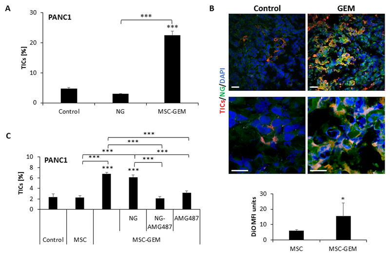
Figure 5. MSC-derived nano-ghosts home to PANC1 tumors in response to gemcitabine therapy:
(A) PANC1 cells were cultured in serum-free medium supplemented as follows: unsupplemented (control); MSC-derived nano-ghosts (NG); and CM derived from gemcitabine-educated MSCs (MSC-GEM). After 3 days, the percentage of TICs in each culture was evaluated by flow cytometry. (B) Eight-to-ten week old SCID mice were orthotopically implanted with PANC1 cells (5x10⁵ cells/mouse) into the pancreas. After 4 weeks, the mice were treated with gemcitabine (500mg/kg) or vehicle control. Twenty-four hours later, mice were intravenously injected with DiO-labelled NGs. After 1 week, mice were sacrificed and tumors were sectioned. Tumor sections were immunostained with antibodies against CD133 to detect TICs (red). Nuclei were stained with DAPI (blue). The mean fluorescence intensity (MFI) of the DiO signal (green) was calculated. Scale bar, 100 µm. (C) PANC1 cells were cultured in serum-free medium supplemented as follows: unsupplemented (control), conditioned medium from untreated MSCs (MSC), CM from gemcitabine-educated MSCs (CM-MSC-GEM) alone or in combination with MSC-derived nano-ghosts (NG), AMG487-loaded NGs (NG-AMG487), or free AMG487 (1µM). After 3 days, the percentage of TICs in each culture was evaluated by flow cytometry. *p<0.05, **p<0.01, ***p<0.001, as assessed by student t-test or one way ANOVA followed by Tukey post-hoc test (when comparing between more than two groups).

