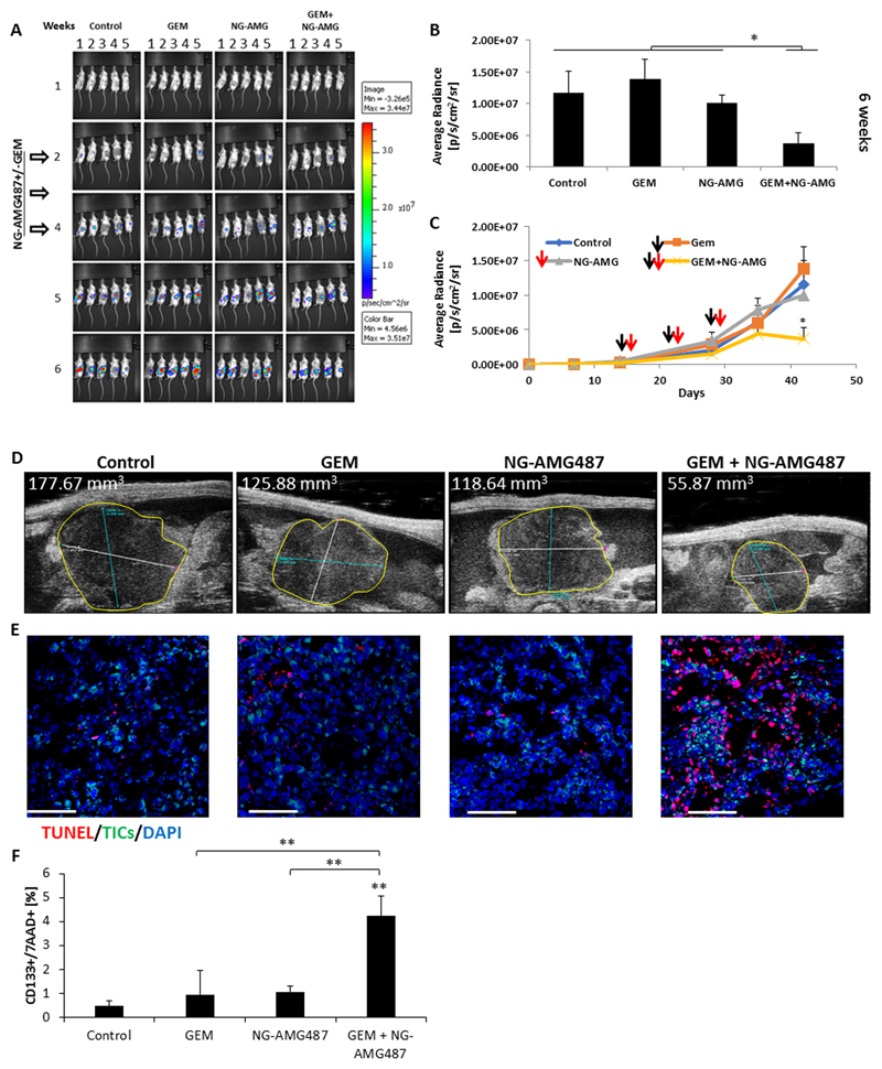
Figure 6. AMG487-loaded nano-ghosts inhibit tumor growth by inducing apoptosis:
(A-D) Eight-to-ten week old SCID mice were orthotopically implanted with PANC1 cells (5x10⁵ cells/mouse). After two weeks, mice were injected with gemcitabine chemotherapy (GEM, 500mg/kg), AMG487-loaded NG (NG-AMG487) or a combination of the two. Gemcitabine was injected weekly one day before the injection of NGs over a 3 week period (white arrows). Note, mouse 3 and 4 in control group week 6 exchanged position. (A) Tumor growth was assessed using the IVIS imaging system. (B-C) Shown are plots of bioluminescence levels. Black arrows represent gemcitabine injections and red arrows represent NG-AMG487 injections. Error bars represent SE. (D) Tumor size at 6 weeks was also analyzed by micro-ultrasound. (E) At end point, tumors were removed. Half of each tumor was sectioned and the other half was prepared as a single cell suspension. Tumor sections were immunostained using antibodies against CD133 to detect TICs (green). Apoptotic cells were detected by TUNEL staining (red). Nuclei were stained with DAPI (blue). Scale bar, 100 µm. (F) The percentage of apoptotic TICs in single cell suspensions was evaluated by flow cytometry. *p<0.05, **p<0.01, ***p<0.001, as assessed by one way ANOVA followed by Tukey post-hoc test (when comparing between more than two groups).

