Fig. 3.
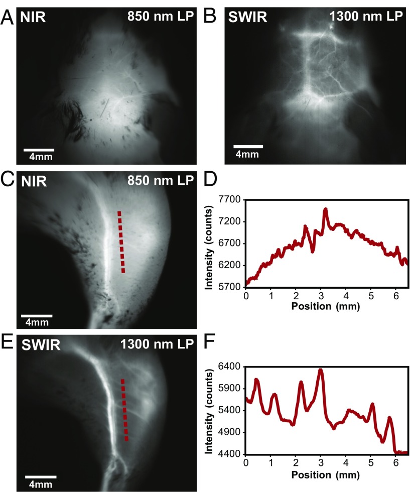
High contrast in vivo SWIR fluorescence imaging using ICG. (A) We noninvasively imaged the brain vasculature of a mouse using ICG contrast and find that the vessels are difficult to resolve through skin and skull using 850-nm long-pass (LP) NIR detection on a silicon camera. (B) Switching to 1,300-nm long-pass SWIR detection on an InGaAs camera greatly improves vessel contrast (Fig. S3 shows contrast quantification). (C) Similarly, only large hind-limb vessels are imaged with good contrast through the skin using NIR detection. (D) The intensity across a line of interest shows insufficient contrast to resolve smaller vessels from background signal. (E) Using 1,300-nm long-pass SWIR detection greatly improves image contrast and (F) resolution of vessels. All images were scaled to the maximum displayable intensities.

