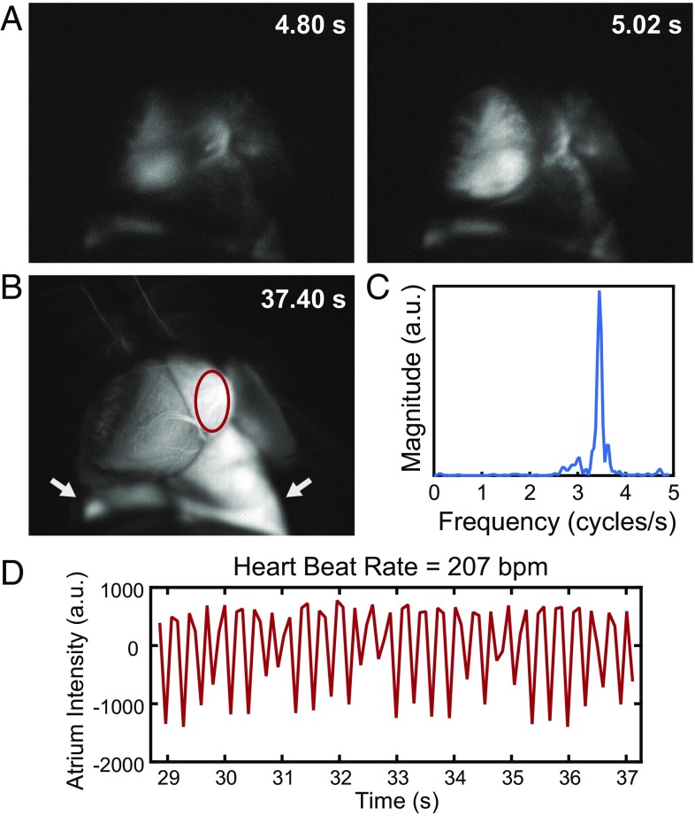
Fig. 4.
High temporal resolution shown through ICG SWIR fluorescence angiography. Intravital SWIR fluorescence angiography was performed in a mouse heart at 9.17 frames per second using ICG for contrast, diffuse 808-nm excitation, and a 1,300-nm long-pass emission filter on an InGaAs SWIR camera (Movie S2). (A) Temporal resolution was sufficiently high to resolve the heartbeat of the mouse. (B) By tracking a region of interest within the atrium of the heart (red circle; lungs are also pictured and indicated with white arrows) and (C) taking the Fourier transform of (D) the intensity fluctuations, the heart rate was determined to be 207 beats per minute for the anesthetized mouse. Fluorescence tracking details and assignment of anatomical structures are in Fig. S5.

