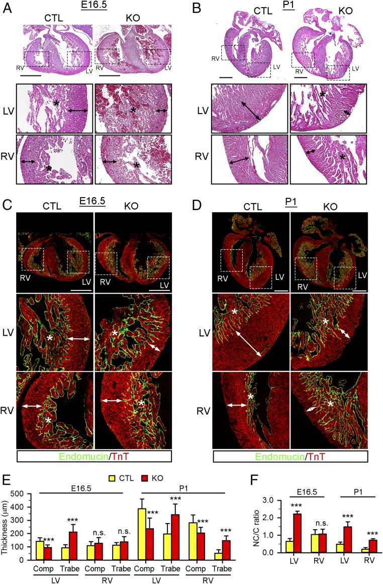
Fig. 2.
NAE1CKO mice displayed ventricular noncompaction. (A and B) Hematoxylin/eosin staining in coronal sections of CTL and NAE1CKO hearts at E16.5 (A) and P1 (B). The Bottom show magnified views of boxed areas in the Top. Double head arrows and asterisks mark the thickness of the compact layer and trabeculae, respectively. (Scale bars: 200 μm.) (C and D) E16.5 (C) and P1 (D) heart sections stained with endomucin (green) and cardiac troponin T (TnT, red) antibodies to delineate the trabecular (covered by endomucin-positive endocardium) and compact layer. (Scale bars: 500 μm.) (E) Quantification of the thickness of the compact (Comp) and trabecular (Trabe) layer in CTL and NAE1CKO hearts. (F) Trabecular layer/compact layer (NC/C) ratio in CTL and NAE1CKO hearts at E16.5 and P1. For morphological analysis, n > 4 hearts per group at each time point were analyzed. Bar graphs are presented as mean + SD. n.s., not significant, ***P < 0.005 versus CTL in unpaired two-tailed t test.

