Fig. 4.
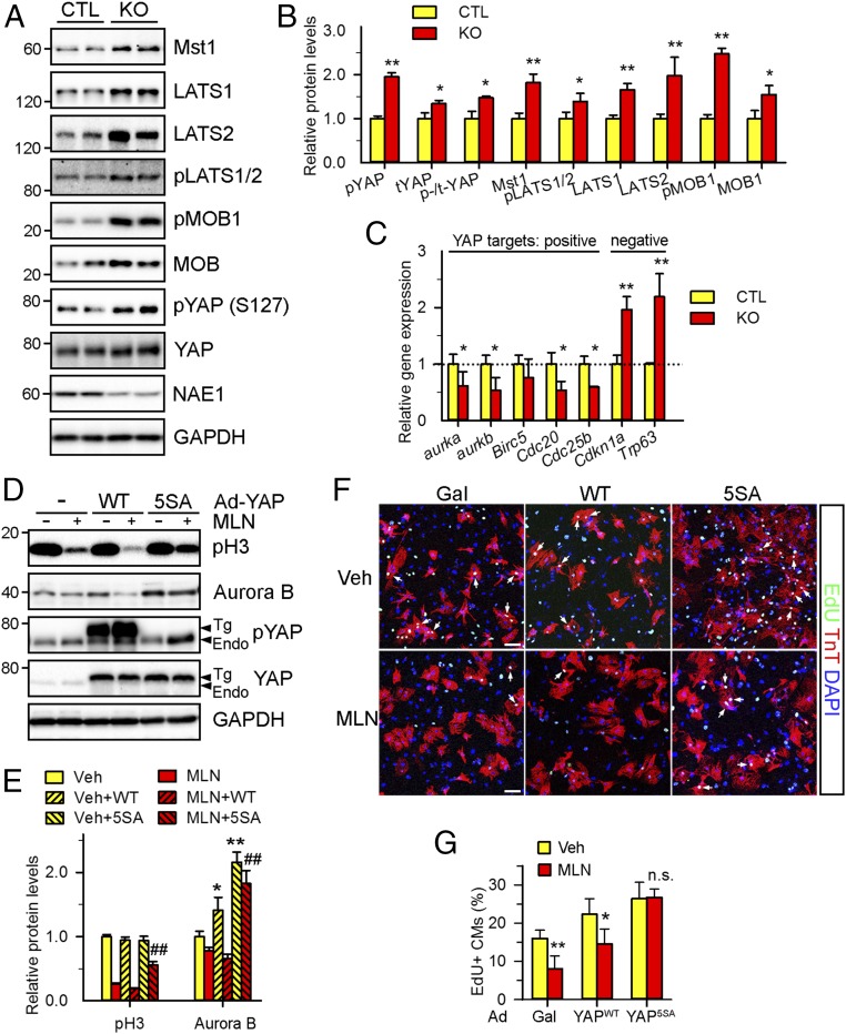
Neddylation controls Hippo-YAP signaling. (A and B) Western blot of the key components of the Hippo-YAP pathway in P1 hearts (A) and the quantification (B). (C) qPCR analysis of the expression of cell cycle genes that are either positively or negatively regulated by YAP in P1 hearts. (D–G) NRVCs were infected with adenoviruses (Ad) expressing WT (YAPWT) or LATS2-insenstive mutant (YAP5SA) form of YAP for 24 h, followed by MLN (1 μM) treatment for additional 24 h. (D and E) Western blots (D) of the indicated proteins and the quantification (E). (F and G) Representative images (F) and the quantification (G) of EdU (green)-labeled cardiomyocytes (arrows). Cardiomyocytes and nuclei labeled by TnT (red) and DAPI (blue), respectively. (Scale bars: 100 μm.) n = 4 fields per group (∼500 cardiomyocytes per field). n.s., not significant; *P < 0.05, **P < 0.01 versus CTL or Veh, ##P < 0.01 versus MLN in unpaired two-tailed t test.

