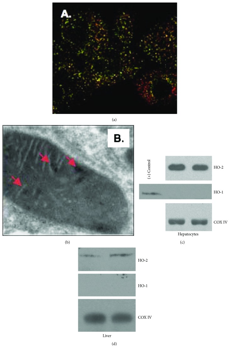
Figure 1.
HO-2 is present within hepatocyte mitochondria under unstressed conditions. (a, b) Microscopy reveals localization of HO-2 within the mitochondria. Immunocytochemistry (a) demonstrates mitochondria (green; MitoTracker) and HO-2 (red) with areas of yellow representing colocalization. Immunoelectron microscopy demonstrates gold particle-labeled localization of HO-2 within hepatocyte mitochondria under normal cell culture conditions (b). (c, d) Western blot of mitochondrial fractions of untreated primary hepatocytes (c) and mouse liver (d) demonstrates HO-2 but not HO-1 expression protein levels under basal cell culture or without treatment. Mitochondrial cytochrome C oxidase subunit 4 (COX IV) is used as a mitochondrial loading control.

