Figure 5.
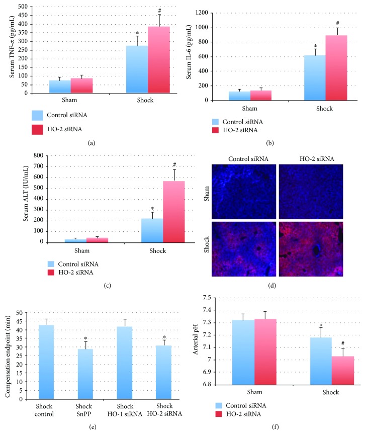
Inhibition of HO-2 exacerbates injury and inflammation in a murine hemorrhagic shock model. (a, b) Knockdown of HO-2 exacerbates hemorrhagic shock-induced serum TNF-α (275 ± 56 control siRNA versus 387 ± 67 HO-2 siRNA) and IL-6 (621 ± 87 control siRNA versus 903 ± 91 HO-2 siRNA). Units are pg/mL; ∗P < 0.05 compared to sham mice and #P < 0.05 compared to shock control siRNA mice. N = 6 mice per group. (c, d) Liver injury and hypoxia were worse in the setting of knockdown of HO-2. Serum ALT increased from 225 ± 59 to 573 ± 102 IU/mL; n = 6 mice per group (c). Hemorrhagic shock also resulted in increased tissue hypoxia as demonstrated by staining for the nitroimidazole EF5, which was also increased by HO-2 siRNA pretreatment (d). (e) Knockdown of HO-2 or nonspecific inhibition of HO activity is associated with earlier decompensation in severe hemorrhagic shock (MAP 20 mmHg). N = 6 mice per group. (f) Arterial pH 30 minutes into severe hemorrhagic shock is decreased compared to control mice (7.32 ± 0.05 versus 7.18 ± 0.08 in shock control siRNA; ∗P < 0.05). This clinical shock parameter is further decreased in HO-2 siRNA-treated mice (7.03 ± 0.06; #P < 0.05 versus shock control siRNA mice). N = 8 mice per group.

