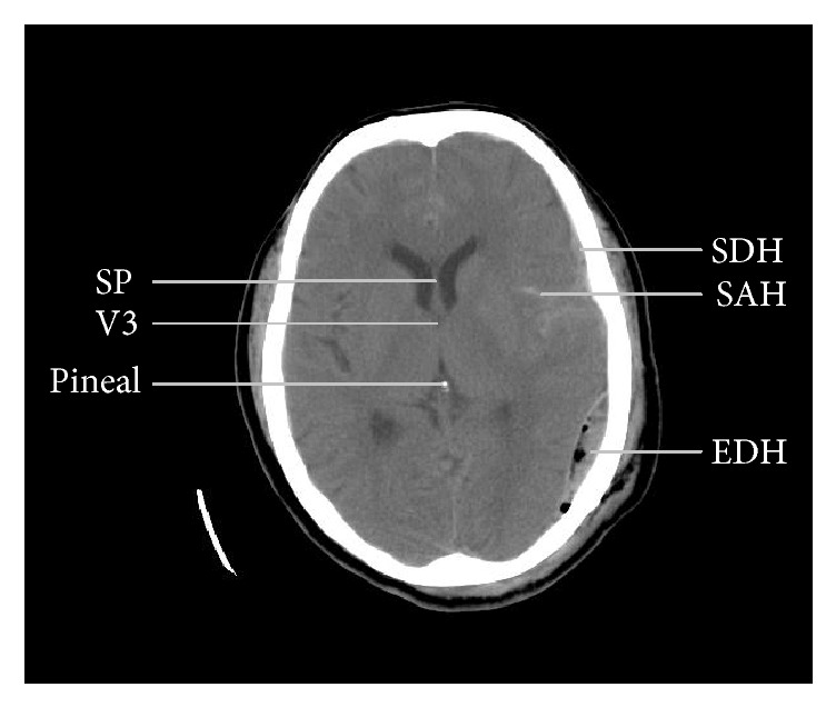
Figure 1.

A computed tomographic image from a patient with traumatic brain injury showing anatomical landmarks used to measure midline shift (2 mm in this image) and different types of intracranial hemorrhage. SP: septum pellucidum, V3: third ventricle (only the most rostral part shown), SDH: subdural hematoma, SAH: subarachnoid hemorrhage, and EDH: epidural hematoma.
