
Figure 2.
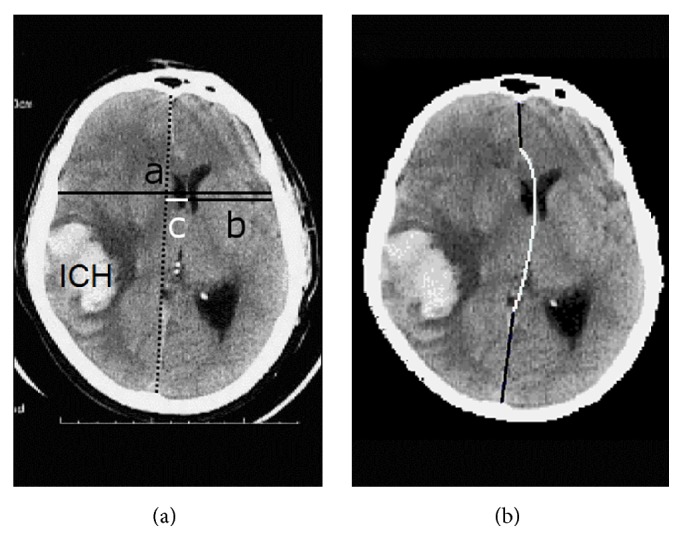
Assessment of midline shift (MLS) on an image of intracerebral hematoma (ICH) compressing the brain. (a) Although determination of the MLS by first measuring the width of the intracranial space (c = a/2 − b) was suggested by the guideline, many neurosurgeons measured it by first drawing the ideal midline (dotted line). (b) Our computational model for the deformed midline included a quadratic Bezier curve (white) between two line segments (black). Adapted from [7].