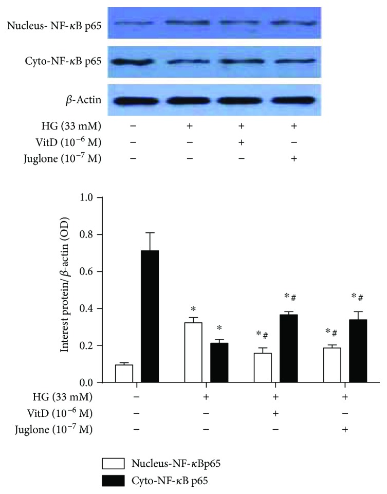
Figure 6.

Effects of vitamin D treatment on NF-κB p65 nuclear translocation in high glucose-cultured HUVECs. HUVECs were inoculated in 6-well plates, cultured with 5% FBS at 70%~80% confluence for 24 h, and then coincubated with high glucose (33 mM) and vitamin D (10−6 M) or Juglone (10−7 M) for 72 h. Total proteins or cytoplasmic/nuclear protein were extracted for immunoblotting analysis. The nuclear and cytoplasmic NF-κB p65 protein levels were expressed as nucleus-NF-κB p65/β-actin and Cyto-NF-κB p65/β-actin, respectively. HG: high glucose (33 mM); nucleus-NF-κB p65: NF-κB p65 in the nuclear; Cyto-NF-κB p65: NF-κB p65 in the cytoplasm; results are presented as mean ± SEM (n = 3); ∗ P < 0.05 versus control; # P < 0.05 versus HG (33 mM).
