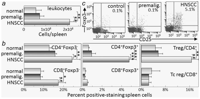
Figure 1.
Increased spleen cellularity, T-cell and Treg levels when premalignant lesions have progressed to oral cancer. Spleen cellularity and T-cell content were assessed in healthy control mice or mice receiving 4NQO treatment for 6 and 16 weeks when premalignant lesions or oral cancer, respectively, were established. Spleen cell mononuclear cell counts were determined (a). Spleen cells were then immunostained for Foxp3 and either CD4 or CD8. Shown are percent spleen cells staining positive as conventional CD4 + Foxp3− or CD8 + Foxp3− cells, regulatory CD4 + Foxp3+ or CD8 + Foxp3+ cells, and the ratio of regulatory to conventional CD4+ or CD8+ cells (b). Also shown are typical dot blots of cells from control mice or mice with either premalignant oral lesions or oral cancer staining as CD25 + Foxp3+ Treg cells (c). * = p <0.05, ** = p <0.01, *** = p <0.001.

