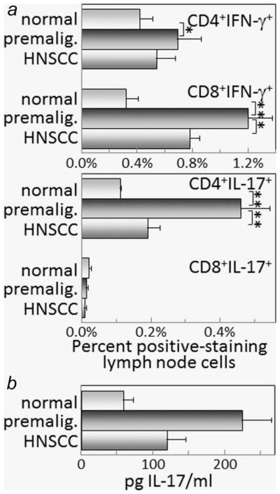
Figure 4.
Increased levels of IFN-γ- and IL-17-staining lymph node cells in mice with premalignant oral lesions. Lymph node cells from healthy control mice, mice with premalignant oral lesions (6 weeks of 4NQO treatment) or mice with oral cancer (16 weeks of 4NQO treatment) were surface immunostained for CD4 or CD8 and stained intracellularly for cytokines. Shown are percentages of spleen cells staining positive for CD4 or CD8 plus either IFN-γ or IL-17 (a). Also shown are levels of IL-17 that are secreted by lymph node cells from control mice or mice with either premalignant oral lesions or oral cancer (b). ** = p <0.01, *** = p <0.001.

