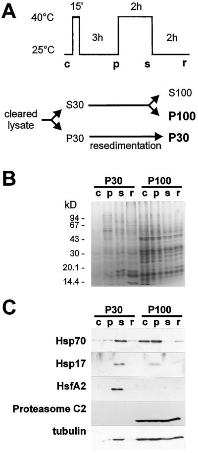
Figure 1.
Proteins in npRNP sediments from control (c), pre-induced (p), stressed (s), and recovered (r) tomato cells. A, Schematic representation of heat-stress treatment and isolation of npRNPs. B, Coomassie blue staining of proteins isolated from different cytoplasmic RNP sediments after SDS-PAGE. C, Immunoblots of RNP sediments probed for HSP70, small HSPs, HsfA2, the proteasome C2 subunit, and tubulin, respectively.

