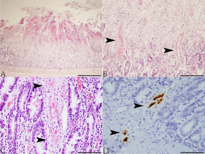
FIG 1.
Photomicrographs of tissues taken from dogs 1 and 2 with typical lesions of CaVV infection. (A) Small intestine, dog 1: severe, acute superficial mucosal necrosis and hemorrhage (H&E stain; scale bar, 850 μm). (B) Small intestine, dog 1: fibrinoid necrosis of the arterial blood vessel walls (arrowheads) at the junction of an area of acute superficial mucosal necrosis and hemorrhage (H&E stain; scale bar, 170 μm). (C) Small intestine, dog 2: fibrinoid necrosis of an arterial blood vessel wall (arrowheads) within the lamina propria (H&E stain; scale bar, 85 μm). (D) Small intestine, dog 2: CaVV nucleic acid signal in endothelial cells of arterial blood vessels (arrowheads) at the junction between the lamina propria and submucosa (ISH; scale bar, 85 μm).

