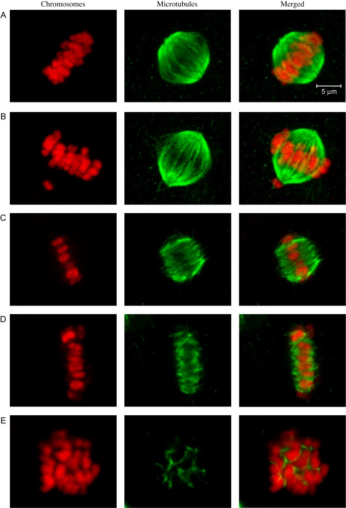
Figure 1.
Representative images of spindle and chromosome classifications in bovine metaphase II oocytes. Chromosomes are shown in red (left panels), microtubules are shown in green (center panels), and merged images are shown in the right panels. (A) Bipolar spindle with aligned chromosomes. (B) Bipolar spindle with a single misaligned chromosome at the lower pole. (C) Bipolar spindle with unfocused poles and aligned chromosomes. (D) Flattened bipolar spindle with extremely broad poles and aligned chromosomes. (E) Non-bipolar spindle with dispersed chromosomes. (The scale applies to all images.)

