Figure 4.
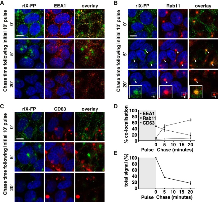
rIX-FP traffics through 293-F FcRn+ cells via a similar route to albumin. A–C, 293-F FcRn+ cells were pulsed with AF488-labeled rIX-FP at pH 5.5 (green) and chased for various times at pH 7. The cells were then fixed and stained with the following organelle specific markers (red): EEA1 for detection of early endosomes (A), Rab11 for recycling endosomes (B), or CD63 for late endosomes/lysosomes (C). The cell nuclei were labeled with Hoechst 33342 (blue). Representative confocal middle sections for each time point are shown. Scale bar, 10 μm. Arrowheads indicate co-localization. Insets show magnified images of selected areas denoted by the white boxes, where the signal for rIX-FP has also been enhanced to highlight co-localization with Rab11. D, the proportion of rIX-FP in each organelle after the 0-, 5-, and 20-min chase time points, as quantified according to “Experimental procedures,” is expressed as % co-localization values. E, the amount of rIX-FP remaining in 293-F FcRn+ cells at each time point is expressed as the relative number of objects/cell, as quantified according to “Experimental procedures.” For D and E, the data represent the means ± S.E. from three independent experiments, where for each experiment, an average value was determined from two to five images (each containing 13 or more cells) for each time point.

