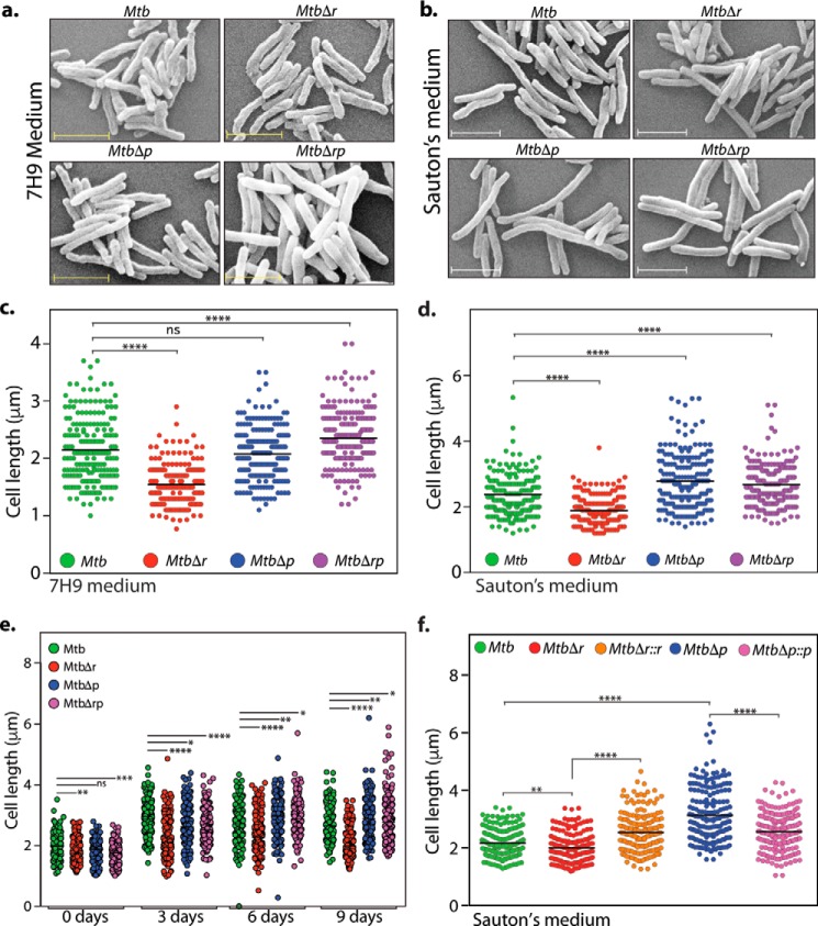
Figure 4.
RodA and PbpA play independent roles in modulating bacterial cell length. a and b, fresh cultures of Mtb, MtbΔr, MtbΔp, and MtbΔrp were seeded at an initial A600 of 0.1 and grown for 6 days at 37 °C at 100 rpm in 7H9 or Sauton's medium followed by fixation. Morphology of the cells was observed through scanning EM at ×20,000 for 7H9 (a) and Sauton's medium (b). Scale bar, 2.0 μm. c, quantification of cell lengths (n ∼200) of Mtb, MtbΔr, MtbΔp, and MtbΔrp strain cells grown in 7H9 medium was performed. Cell lengths were measured independently using Smart Tiff software and plotted by a scatter dot plot with mean values using GraphPad Prism version 6. Mean cell lengths obtained were as follows: Mtb, 2.1 μm; MtbΔr, 1.5 μm; MtbΔp, 2.0 μm; MtbΔrp, 2.3 μm. The experiments were biologically and technically repeated twice. Cell lengths were analyzed using GraphPad Prism version 6, and statistical analysis was performed using a one-way ANOVA test. ****, p < 0.0001; ***, p < 0.001; ns, not significant. d, quantification of cell lengths in Mtb, MtbΔr, MtbΔp, and MtbΔrp strain (n ∼200) cells grown in Sauton's medium was performed, and mean cell lengths obtained were as follows: Mtb, 2.3 μm; MtbΔr, 1.8 μm; MtbΔp, 2.7 μm; MtbΔrp, 2.6 μm. The experiments were biologically and technically repeated twice. Cell lengths were measured independently using Smart Tiff software and plotted by a scattered dot plot with mean values using GraphPad Prism version 6, and the statistical analysis was performed with a one-way ANOVA test. ****, p < 0.0001; ***, p < 0.001. e, fresh cultures of Mtb, MtbΔr, MtbΔp, and MtbΔrp were seeded at an initial A600 of 0.1 and grown at 37 °C at 100 rpm in Sauton's medium followed by fixation at different time points (days 0, 3, 6, and 9) of growth, and cell lengths were measured as described above. Mean cell lengths obtained for different time points were as follows: day 0: Mtb, 1.98 μm; MtbΔr, 1.80 μm; MtbΔp, 1.84 μm; and MtbΔrp, 1.76 μm; day 3: Mtb, 2.87 μm; MtbΔr, 2.26 μm; MtbΔp, 2.69 μm; and MtbΔrp, 2.62 μm; day 6: Mtb, 2.72 μm; MtbΔr, 2.32 μm; MtbΔp, 2.92 μm; and MtbΔrp, 2.90 μm; and day 9: Mtb, 2.71 μm; MtbΔr, 2.0 μm; MtbΔp, 2.91 μm; and MtbΔrp, 2.88 μm. Cell lengths were analyzed using GraphPad Prism version 6, and statistical analysis was performed using a one-way ANOVA test. ****, p < 0.0001; ***, p < 0.001; **, p < 0.01; ns, not significant. f, fresh cultures of Mtb, MtbΔr, MtbΔr::r, MtbΔp, or MtbΔp::p were seeded at an initial A600 of 0.1 in Sauton's medium and continued to grow for 6 days in the presence of 0.1 μm IVN, and cells were fixed and processed for SEM. Cell lengths were observed through SEM at ×20,000. Cell lengths were measured as described above. Obtained mean cell lengths were as follows: Mtb, 2.1 μm; MtbΔr, 1.9 μm; MtbΔr::r, 2.5 μm; MtbΔp, 3.1 μm; MtbΔp::p, 2.5 μm. Statistical analysis was performed using a two-way ANOVA test; ****, p < 0.0001; **, p < 0.01.

