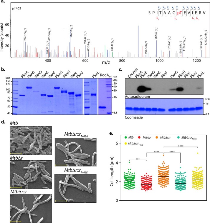
Figure 8.
RodA is phosphorylated on Thr-463 residue. a, MS/MS spectrum of precursor m/z: 761.88025 (+2) and MH+: 1522.75322 Da, of the phosphopeptide SPITAAGpTEVIERV (where pT represents phosphothreonine). The location of Thr-463 was evident by the presence of ion series containing y6, y7, b3, b7, and y8–11 in the spectra. b, Coomassie-stained purified MBP-STPKs and His-FLAG-RodA(411–469). c, an in vitro kinase assay was performed with 10 pmol of MBP-STPKs and 312 pmol of His-FLAG-RodA(411–469). Samples were resolved on 15% SDS-PAGE, stained with Coomassie (bottom), and autoradiographed (top). d, fresh cultures of Mtb, MtbΔr, MtbΔr::r, MtbΔr::rT463A, or MtbΔr::rT463E were seeded at an initial A600 of 0.1 in 7H9 medium and continued to grow for 6 days in the presence of 100 ng of anhydrotetracycline, followed by fixation. SEM was performed to analyze the morphology of the cells (×20,000). Scale bar, 2 μm. e, cell lengths of ∼200 individual cells for each sample were quantified. Mean cell lengths for the samples were 2.1 μm (Mtb), 1.6 μm (MtbΔr), 2.5 μm (MtbΔr::r), 1.8 μm (MtbΔr::rT463A), and 2.2 μm (MtbΔr::rT463E). Similar results were obtained in a biological replicate. Statistical analysis was performed using a one-way ANOVA test; ****, p < 0.0001; ***, p < 0.001.

