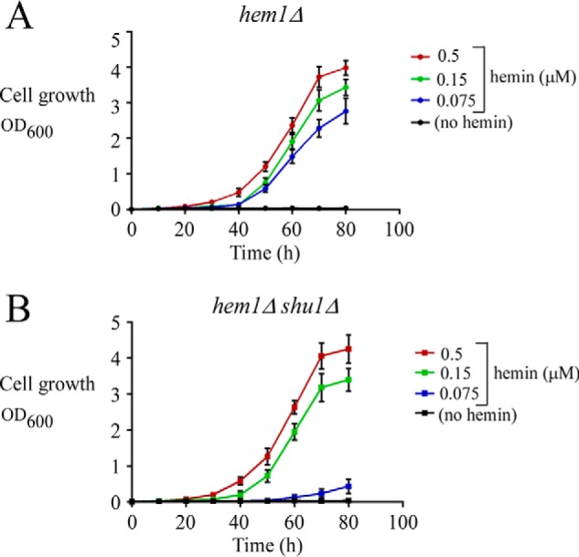
Figure 1.

Deletion of shu1+ leads to poor growth in the presence of low hemin concentration (0.075 μm) but does not compromise cell growth with higher hemin concentrations (0.15 and 0.5 μm). A, growth of hem1Δ cells was assessed in ALA-free medium that was left untreated (no hemin; black) or supplemented with exogenous hemin. Hemin color codes are as follows. Blue, 0.075 μm; green, 0.15 μm; red, 0.5 μm. B, growth of hem1Δ shu1Δ cells was assessed under the same conditions as described for A. Values are represented as the averages ± S.D. (error bars) of a minimum of three independent experiments.
