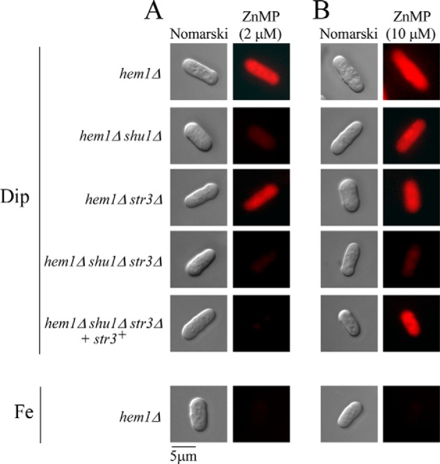
Figure 4.

Deletion of shu1+ and str3+ leads to defects in the cellular assimilation of ZnMP, albeit in a different concentration threshold. The indicated strains were precultured in the presence of Dip (50 μm) or FeCl3 (100 μm) and ALA (200 μm). Cells were transferred in ALA-free medium containing Dip (250 μm) or FeCl3 (100 μm) for 3 h. In the final 90 min of treatment, ZnMP was added at the indicated concentration (2 μm (A) or 10 μm (B)). A hem1Δ shu1Δ str3Δ triple mutant strain in which a WT copy of the str3+ gene was reintegrated was cultured in an identical manner. Cells were analyzed by fluorescence microscopy for accumulation of fluorescent ZnMP (right). Nomarski optics (left) was used to examine cell morphology. Results of microscopy are representative of five independent experiments.
