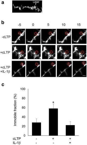
Fig. 4.

IL-1β treatment decreased the cLTP-induced F-actin stabilization. FRAP analyses were performed using hippocampal neurons at 16–18 DIV that were transfected with Lifeact-GFP. a Illustration of FRAP setting. b Representative fluorescence images show spines of control group (-cLTP, top panel), cLTP group (middle panel), and cLTP+IL-1β group (bottom) during FRAP. Scale bar, 2 μm c Analysis of immobile fractions from data obtained in b. The mobile fraction and the immobile fraction (fi) were calculated as described in the “Methods” section. Data are mean ± SEM (n = 4, (*p < 0.05, ANOVA)
