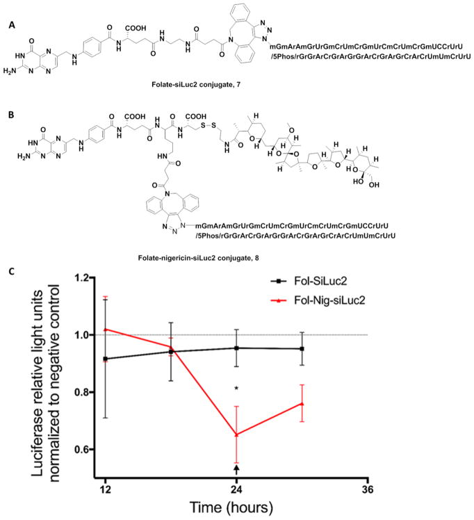
Figure 8.
Nigericin-mediated release of Folate-conjugated siLuc2. (A) Structure of Fol-siLuc2 7. (B) Structure of Fol-nigericin-siLuc2 conjugate 8. (C) Luciferace2 enzyme activity in MDA-MB-231 cells as a function of time after treatment with either folate-siLuc2 or folate-nigericin-siLuc2 conjugate. Luciferase activity levels were normalized to a negative control (a scrambled siRNA). Mean ± S.D., technical replicates = 3, n = 3, * P < 0.05. The arrow indicates replacement of media with a new dose of folate conjugates (50 nM). Fol-SiLuc2: Folate-siLuc2; Fol-Nig-SiLuc2 conjugate: Folate-nigericin-siLuc2 conjugate. The gene-knockdown experiment indicated a rapid reduction in luciferase activity in Fol-nigericn-siLuc2 treated cells as soon as 18 h post treatment and reaches 30–35% after 24 h.

