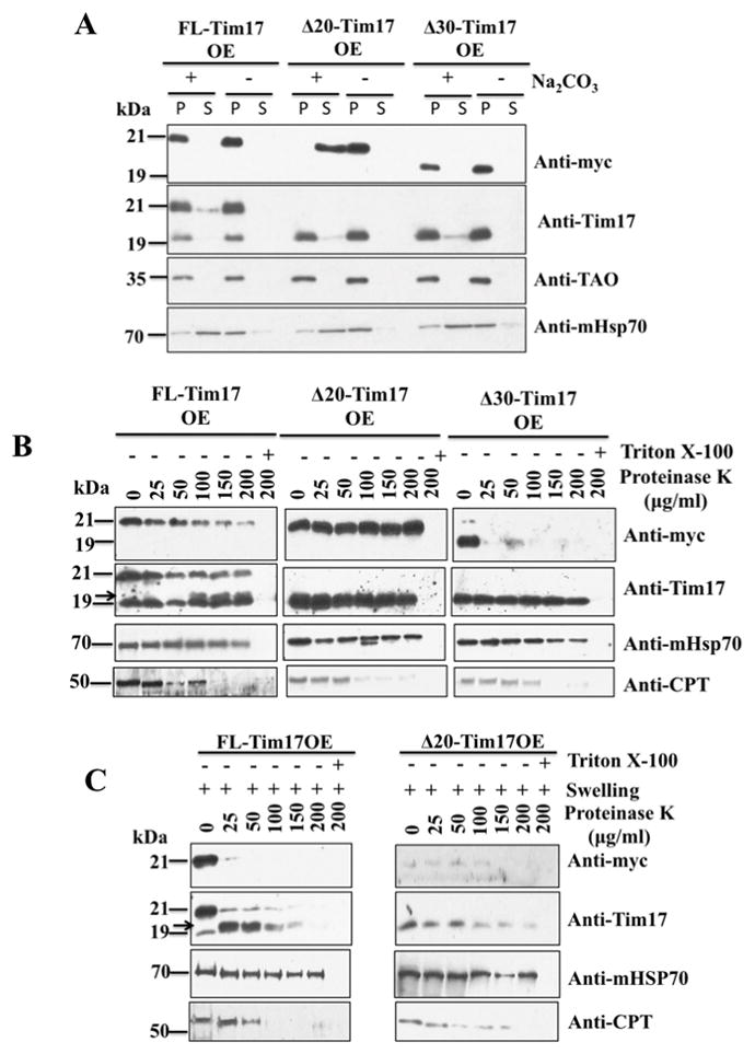
Fig. 4.

Sub-mitochondrial localization of the N-terminal deletion mutants of TbTim17. (A) Alkali extraction of the mitochondria isolated from FL-Tim17OE, Δ20-Tim17OE, and Δ30-Tim17OE after growing in the presence of doxycycline for 48 h. Proteins from equal volume of the supernatant (lane S) and pelleted (lane P) fractions were analyzed by immunoblotting using anti-myc, -Tim17, -TAO, and –mHSP70 antibodies. (B) Mitochondria isolated from the FL-Tim17OE, Δ20-Tim17OE, and Δ30-Tim17OE T. brucei grown for 48 h in the presence of doxycycline were treated with various concentration (0–200 μg/ml) of proteinase K as described in the materials and Methods. Proteins were analyzed by immunoblotting using anti-myc, -Tim17, mHSP70, and –CPT antibodies. (C) Mitochondria from the FL-Tim17OE and Δ20-Tim17OE T. brucei were swelled in hypotonic buffer to rupture the mitochondrial outer membrane. Mitoplasts were recovered by centrifugation and treated with various concentrations (0–200 μg/ml) of proteinase K as described above. Proteins were analyzed by immunoblot analysis using anti-myc, -Tim17, -mHSP70, and -CPT antibodies. Arrow shows a ~19 kDa cleaved product of FL-Tim17-2X-myc.
